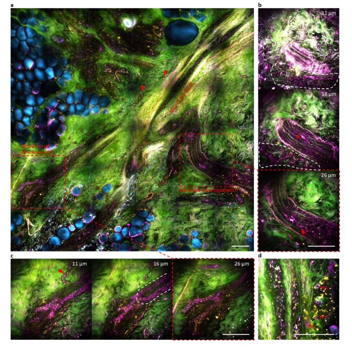
Fig. 6.
Wide-field and selective volumetric tetra-modal imaging of rejuvenated magenta-colored field cancerization by chemotherapeutically eliminating multifocal primary tumors in a cancer patient (Subject #393). (a) Wide-field tetra-modal image of the peri-tumoral field with root-like mammary ducts, with two regions of interest subjective to selective volumetric tetra-modal imaging (broken boxes). (b) Selective volumetric tetra-modal images at three depths showing branched mammary tubulogenesis with luminal epithelial cells (arrowhead) and myoepithelial cells (arrow), which intertwines with developing capillaries surrounded by magenta-colored stromal cells (region of white broken curve) (Visualization 3 (7.1MB, avi) ). (c) Selective volumetric tetra-modal images at three depths showing blood vessel development of a capillary (arrowhead) and a connected sprouting vessel (region of white broken curve) (Visualization 4 (8.5MB, avi) ). (d) main branch of mammary ducts with peripheral magenta-colored stromal cells (arrows) different from yellow-colored stromal cells. Scale bars: 100 µm.

