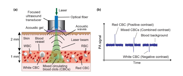
Fig. 1.
The principle of PA detection of red, white and mixed CBCs producing positive, negative and combined PA contrast peaks in blood background, respectively. (a) Schematic of acoustic resolution PAFC diagnostic platform using a high pulse-repetition-rate laser and focused ultrasound transducer with a central hole to deliver laser light through a fiber. (b) PA trace showing PA peaks with positive (red CBC), negative (white CBC) and combined (mixed CBCs) contrast in the blood background.

