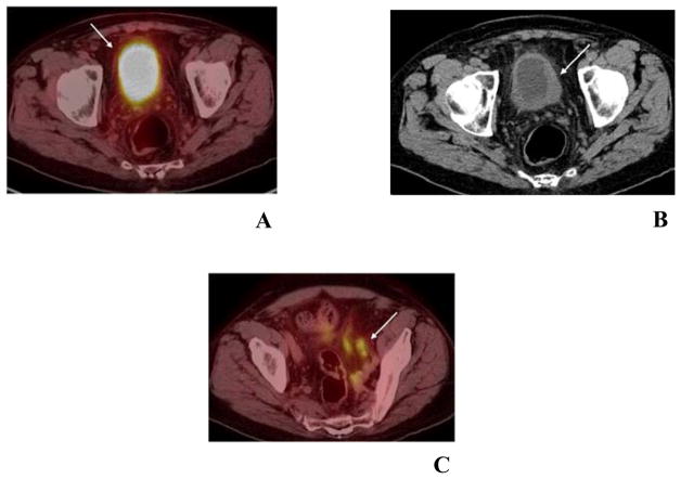
Fig. 4.
78-year-old man with large papillary tumor on left lateral wall and high grade, nonpapillary tumor involving the left trigone just lateral to left UO and posterior left bladder neck on cystourethroscopy.
Due to concentration of 18F-FDG in the urinary bladder, any increased activity of the thickened bladder wall (arrow) cannot be determined on the PET/CT image of the pelvis (A). Asymmetric thickening of the left urinary bladder wall (arrow), on the pelvis CT part of PET/CT imaging (B) and it likely corresponds to known bladder malignancy. Along the left pelvic sidewall there is a 2.4 × 1.3 cm hypermetabolic lymph node with a maximum SUV of 6.17 (C).

