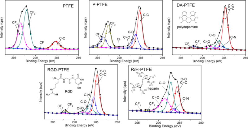
Figure 2.

Gauss-fitted C1s high-resolution scans of PTFE, P-PTFE, DA-PTFE, RGD-PTFE, and R/H-PTFE showing the composition of different carbon bonds. Statistical data is listed in Table S2 in the Supporting Information. The insets show the chemical structure of polydopamine, RGD, and heparin.
