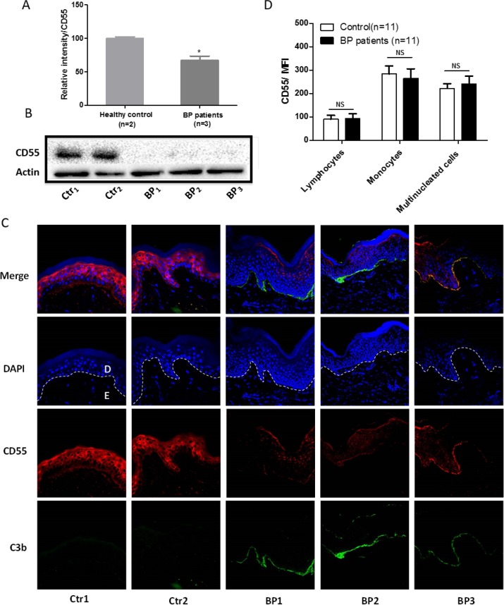
Figure 1. CD55 downregulation in the lesional skin of patients with bullous pemphigoid.
CD55 expression in bullous pemphigoid skin sections was examined by qRT-PCR (A) and western blotting (B). (C) Normal skin sections and lesional skin specimens from patients with bullous pemphigoid were subjected to immunofluorescent staining to measure CD55 expression in epidermal keratinocytes. (D) Flow cytometric analysis of CD55 expression on peripheral blood cells. Results are presented as the means ± SEM from 11 independent observations. D, dermal; E, epidermal. NS, no significant difference between controls and bullous pemphigoid patients. DAPI-stained nuclei can be seen in blue. Scale bar, 100 nm.

