Figure 2.

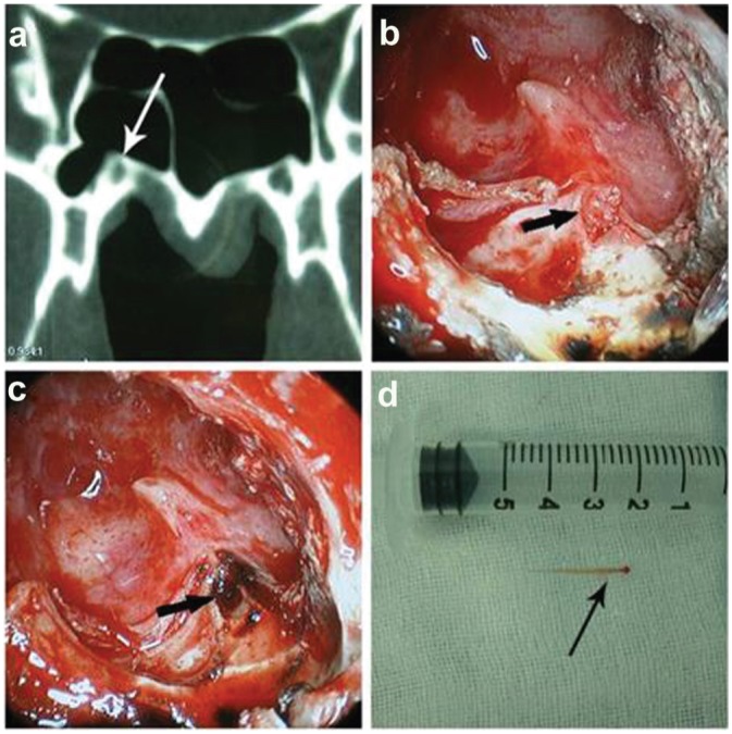
Computed tomography (CT) scan–guided surgery. Both CT scan of the vidian canal (a) and endoscopic view of the vidian nerve during surgery (b, nerve exposed; c, after cautery) with a section of resected vidian nerve (d, gross photograph).

Computed tomography (CT) scan–guided surgery. Both CT scan of the vidian canal (a) and endoscopic view of the vidian nerve during surgery (b, nerve exposed; c, after cautery) with a section of resected vidian nerve (d, gross photograph).