
Figure 5.
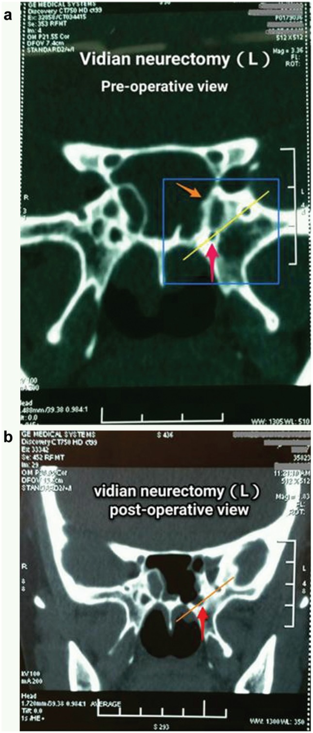
Computed tomography (CT) scan of vidian canal with pre- (a) and postoperative (b) comparison. Bony part of vidian canal indicated by red arrows in Figure 5a has been removed, as shown in Figure 5b . Bony septum referred to by orange arrow has been removed to expose the vidian nerve canal, as shown in Figure 5b .