
Fig. 2.
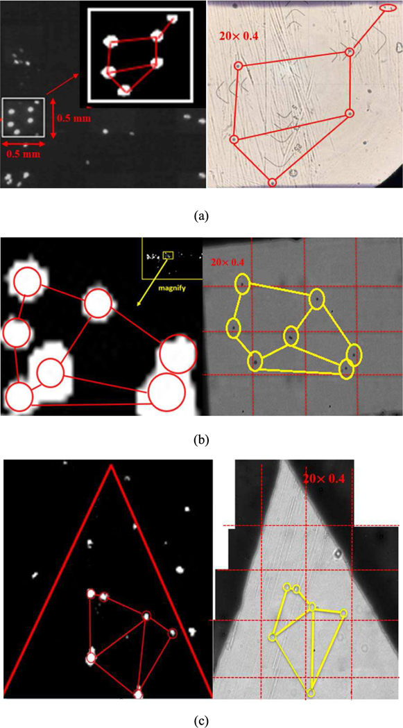
Comparisions of particle distribution pattern between the CMOS imager image and the optical microscope image mosiac at 20 times optical magnification and 0.4 numerical aperture. Size of particles used are (a) 10 µm, (b) 5 µm, and (c) 0.5 µm.
Official websites use .gov
A
.gov website belongs to an official
government organization in the United States.
Secure .gov websites use HTTPS
A lock (
) or https:// means you've safely
connected to the .gov website. Share sensitive
information only on official, secure websites.
Comparisions of particle distribution pattern between the CMOS imager image and the optical microscope image mosiac at 20 times optical magnification and 0.4 numerical aperture. Size of particles used are (a) 10 µm, (b) 5 µm, and (c) 0.5 µm.