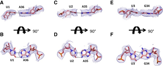
FIGURE 3.
X-ray crystallography structures of the decoding mRNA-ASL minihelix. (A) Simple Fo-Fc omit maps of the decoding complex individual base pair mRNA(U1)-ASL(A36) contoured at the 3σ level, colored in gray and shown at 3 Å. (B) Same as A rotated around x-axis by 90°. (C) Simple Fo-Fc omit maps of the decoding complex individual base pair mRNA(U2)-ASL(A35) contoured at the 3σ level, colored in gray and shown at 3 Å. (D) Same as C rotated around x-axis by 90°. (E) Simple Fo-Fc omit maps of the decoding complex individual base pair mRNA(U3)-ASL(G34) contoured at the 3σ level, colored in gray and shown at 3 Å. (F) Same as E rotated around the x-axis by 90°.

