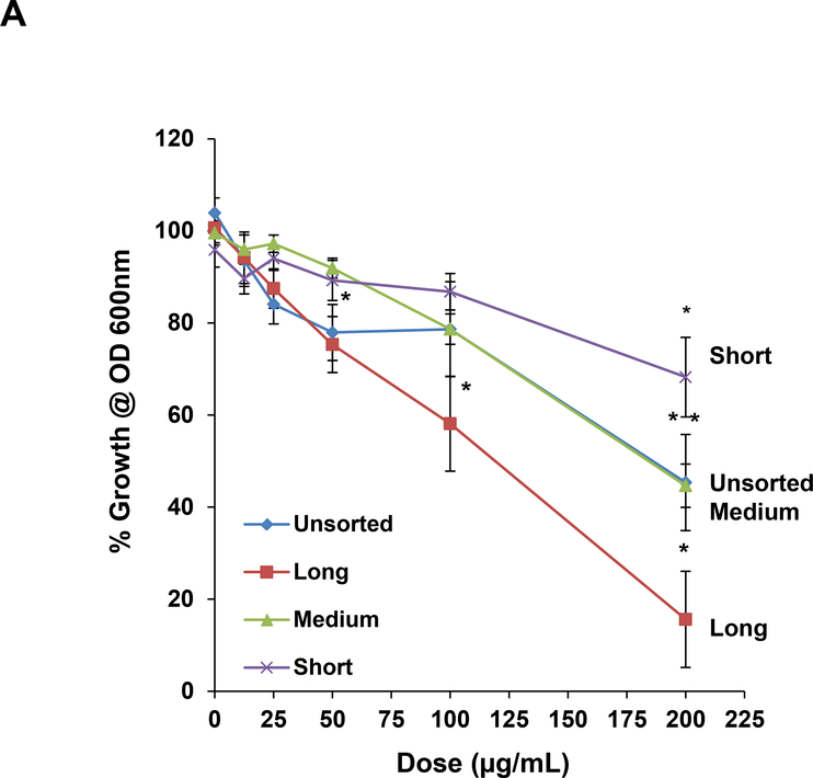
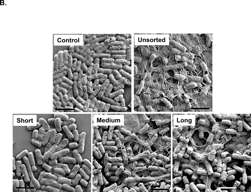
Figure 4: Assessment of the antibacterial effects of length-sorted SWNCTs in E. coli.
(A) E. coli cell growth was assessed by optical absorbance (OD 600 nm) in a plate reader (Spectro Max M5e, Molecular Devices, Sunnyvale, CA, USA). The cultures were incubated with 12.5, 25, 50, 100 and 200 μg/mL SWNCTs for 24 h in regular LB medium. (B) Scanning electron microscopy (SEM) was used to assess morphological changes in E. coli exposed to sorted and unsorted tubes at 200 μg/mL for 24 h. After fixation and dehydration, the cells were imaged under a JEOL JSM-67 field emission scanning electron microscope at 10 kV. The scale bars represent 2 μm in length.


