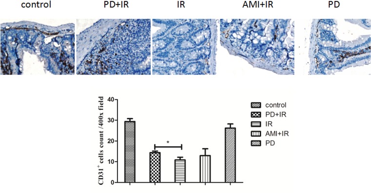
Figure 5. Immunohistochemical staining of small intestine with CD31 antibody.
*, P < 0.05. 400× magnification. Blood vessels were identified in vascular endothelial cells/cell clusters (brown) and were distinct from the background. A total of 10 fields of view (400×) were randomly selected in each mouse to determine MVD. PD dosage, 25 mg/kg.

