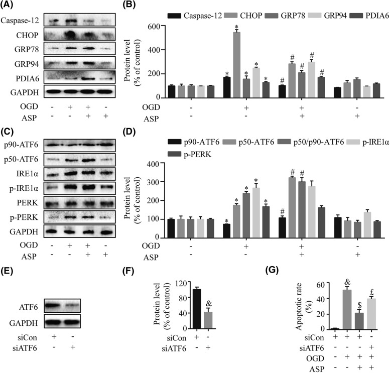
Figure 3. ASP relieves OGD-induced ER stress by activating the ATF6 branch.
The expression levels of the ER stress-induced pro-apoptosis mediator (CHOP and Caspase-12) and anti-apoptotic molecules (GRP78, GRP94, and PDIA6) were determined by Western blot (A) and densitometry (B) analyses. The expressions of three transducers in the UPR including ATF6, IRE1α, and PERK were measured by Western blot (C) and densitometry (D) analyses. (E–G) H9c2 cells were transfected with siCon and specific siATF6, and then stimulated with OGD for 4 h with or without 50 µg/ml ASP pretreatment. Western blot (E) and densitometry (F) analyses demonstrated a significant knockdown of ATF6 in H9c2 cells after treatment with siATF6. (G) The flow cytometric analysis showed the beneficial effect of ASP was blunted after transfection with siATF6. *P<0.05 compared with the control group; #P<0.05 compared with the OGD-treated group; &P<0.05 compared with the siCon group; $P<0.05 compared with the siCon + OGD group; £P<0.05 compared with the siCon + OGD + ASP group.

