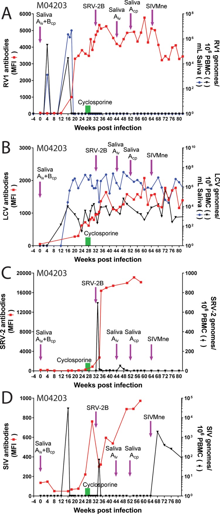
Fig 5. Experimental rhadinovirus infection in juvenile macaque M04203.

The naïve juvenile pig-tailed macaque M04203 was inoculated intravenously (i.v.) with diluted filtered saliva from the 01019 cynomolgus macaque donor (Saliva “A”) containing high levels of RFHVMf and low levels of MfaLCV and was inoculated into the cheek pouches with whole saliva from pig-tailed macaque A02241 (Saliva “B”), which had high levels of RFHVMn, as indicated in the text. M04203 received a second iv inoculation of saliva from 01019 (Saliva “A”) 44 weeks after the initial infection. Subsequently, whole saliva from 01019 (Saliva “A”) was administered to the cheek pouch at week 52. To activate rhadinovirus infections, M04203 was treated with daily injections of cyclosporine for 3 weeks (week 26–29) to induce immune suppression, as indicated (green bar). The animal was subsequently infected with SRV-2B and SIVMne, which have previously been associated with rhadinovirus activation and associated pathologies (see text). Saliva and blood were collected weekly and/or bi-weekly. A) The RV1 genome levels were determined using the RV1 qPCR assay and the RV1 antibody levels were determined using the RV1 Luminex assay. B) the LCV genome levels were determined using the LCV qPCR assay and the LCV antibody levels were determined using the LCV Luminex assay. C) The SRV2 cDNA levels were determined using the SRV2 qPCR assay and the SRV2 antibody levels were determined using the SRV2 Luminex assay. D) The SIV cDNA levels were determined using the SIV qPCR assay and the SIV antibody levels were determined using the SIV Luminex assay.
