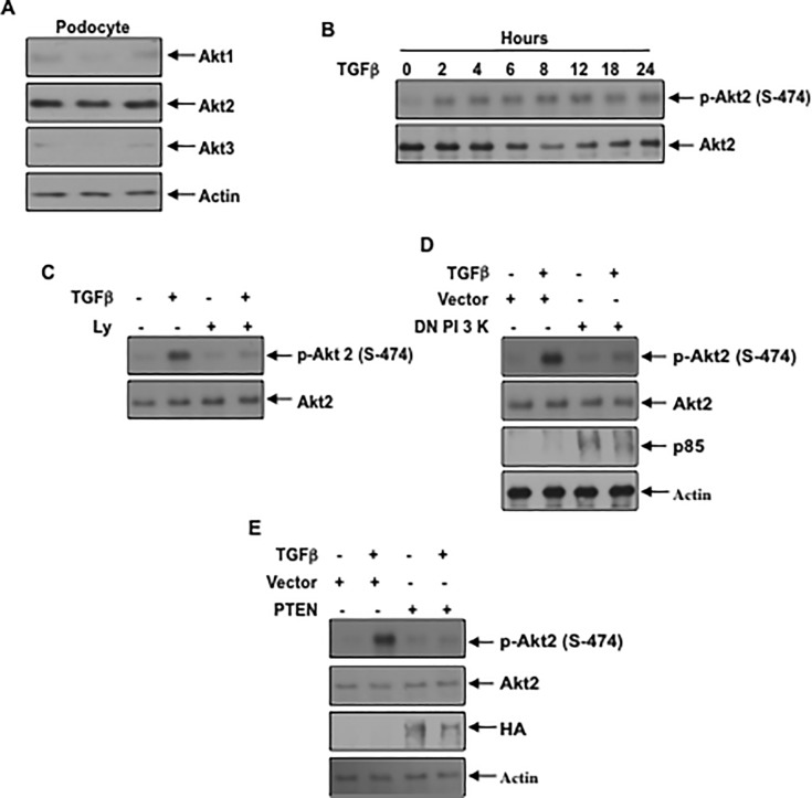
Fig 5. Akt2 is abundantly expressed in podocytes and PI 3 kinase regulates its activation by TGFβ (A) Podocyte lysates from independent dishes were immunoblotted with isoform-specific Akt1, Akt2, Akt3 and actin antibodies.
(B) TGFβ increases phosphorylation of Akt2 in a time-dependent manner. Serum-starved podocytes were incubated with 2 ng/ml TGFβ for the indicated periods of time. The cell lysates were immunoblotted with the phospho-Akt (Ser-474) specific antibody. As a control Akt2 specific antibody was used. (C) PI 3 kinase inhibition blocks TGFβ stimulated phosphorylation of Akt2. Serum-starved podocytes were incubated with 25 micromolar Ly before treatment with TGFβ. The cell lysates were immunoblotted with the indicated antibodies. (D and E) Podocytes were transfected with dominant negative p85 subunit of PI 3 kinase (panel D) or PTEN (panel E). The transfected cells were serum starved and incubated with TGFβ The cell lysates were immunoblotted with the indicated antibodies. Representative of 3 and 4 independent experiments is shown for panels B and C, D, E. The quantification is shown in the S7 Fig.

