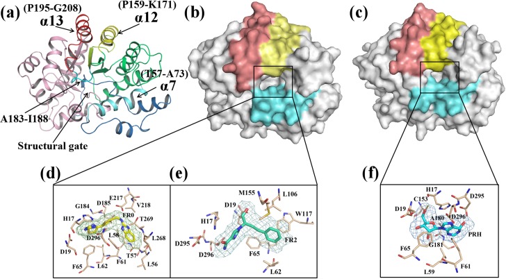
Fig 1. Overview of adenosine deaminase (ADA, PDB code: 1VFL) and the three ligands.
(a) ADA structure. The α12 helix (P159-K171) is colored yellow; the α13 helix is colored red; and the structural gate is colored cyan, which consists of the α7 helix (T57-A73) and residues A183 to I188. (b) The binding pocket of ADA with an open form. (c) The binding pocket of ADA with a closed form. (d) The ligand FR0 (obtained from PDB: 1DNV). (e) The ligand FR2 (obtained from PDB: 1NDW) and (f) the ligand PRH (obtained from PDB: 1KRM) are denoted by sticks and mesh. The surrounding residues are depicted by light orange sticks.

