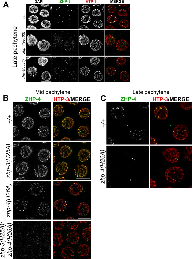
Fig 4. The RING finger domain of ZHP-4 is required for localization of ZHP-3/4.
(A) Representative images of late pachytene nuclei of wild types, zhp-4(vv103) and zhp-4(vv96) mutants stained with α-ZHP-3 (green) and α-HTP-3 (red). In wild-type, ZHP-3 restricted to ~6 foci designating crossover sites per nucleus, while in zhp-4(vv103) mutants, no ZHP-3 SC localization or foci could be detected). In zhp-4(vv96) mutants, ZHP-3 foci and occasionally small stretches were detected and colocalized with synapsed HTP-3 marked axes. (B) Representative images of mid-pachytene nuclei of the indicated genotypes stained for α-ZHP-4 (green) and α-HTP-3 (red). Both wild-type and zhp-3(H25A) mutant nuclei show ZHP-4 localization with synapsed chromosome axes. In zhp-4(H26A) mutant nuclei only a punctate staining of ZHP-4 is detectable; occasional small brighter foci that colocalize with HTP-3 amongst a larger population of weak dimmer foci distributed throughout the nucleus. In zhp-3(H25A);zhp-4(H26A) double mutants ZHP-4 is not detectable above background levels. (C) Representative images of late pachytene nuclei stained with α-ZHP-4 (green) and α-HTP-3 (red). Wild-type nuclei largely display ~ 6 ZHP-4 foci on chromosome axes and 1–3 similarly-sized bright foci can be detected in zhp-4(H26A) RING mutants. Scale bars 5 μm.

