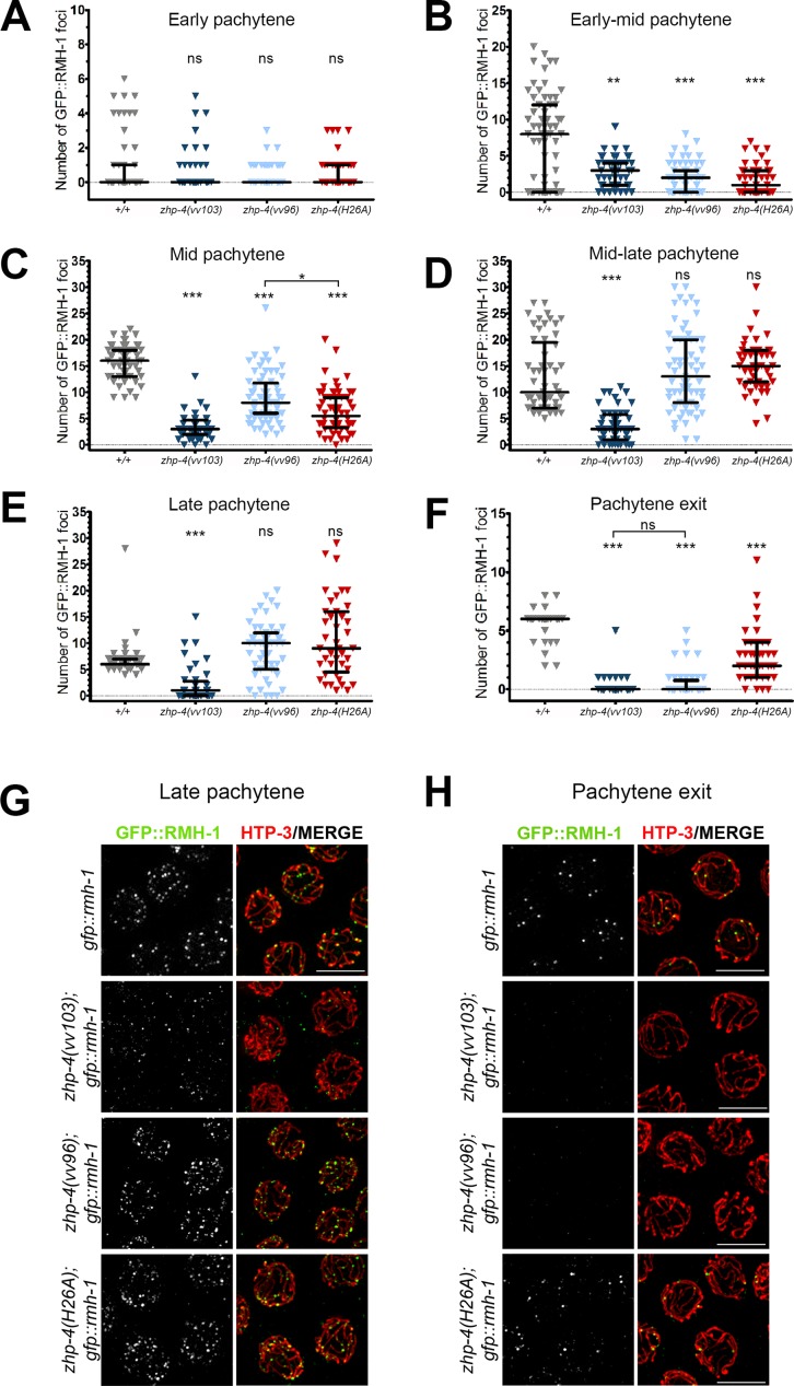
Fig 6. ZHP-4 is required to stabilize RMH-1 at early and late recombination intermediates.
GFP::RMH-1 was quantified in the germline nuclei of animals of the indicated genotypes and the numbers binned into six equal zones encompassing the pachytene region: early (A), early-mid (B), mid (C), mid-late (D), late pachytene (E) and pachytene exit (F). All nuclear foci were quantitated. In wild types, RMH-1 appearance and kinetics are similar to a previous report [12]. Three gonads were scored for each genotype and the data are represented as individual points grouped by genotype and plotted on each Y axis; black bars represent the median +/- interquartile range and Kruskal-Wallis tests and multiple pairwise comparisons were used to assess significant differences among mutants versus wild types (ns = not significant, ** p<0.01, *** p<0.001). (G,H) Immunolocalization of GFP::RMH-1 (green) and HTP-3 (red) in late pachytene nuclei of the indicated genotypes detects a few small foci in zhp-4(vv103) mutants and multiple brighter foci in zhp-4(vv96) and zhp-4(H26A) mutant nuclei. At pachytene exit, no late RMH-1 foci were detectable in zhp-4(vv103) and zhp-4(vv96) mutants, while 2–4 foci with sizes similar to wild-type foci could be detected on HTP-3-marked tracks in the RING mutant. Scale bars 5 μm.

