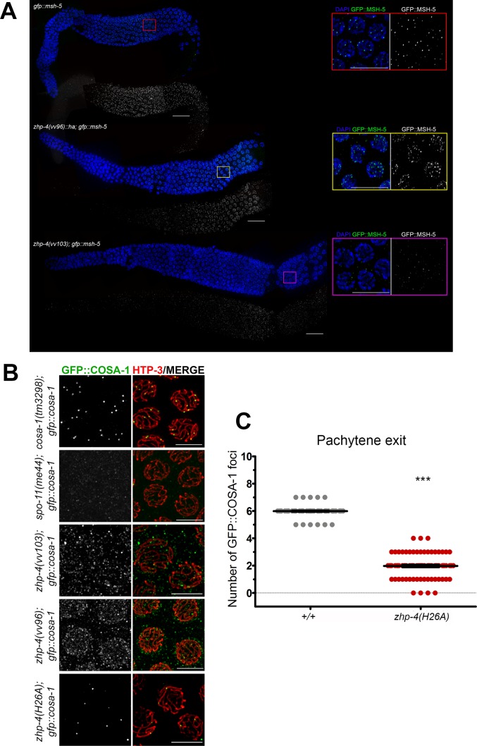
Fig 7. zhp-4 mutants are defective in acquiring late crossover markers.
(A) Whole gonad immunostaining with DAPI (blue) and α-GFP (green) antibody marking GFP::MSH-5 in the indicated genotypes. Pachytene progression proceeds from left to right and insets show magnifications of late pachytene stage nuclei from the corresponding genotype. In wild types, abundant small MSH-5 foci appearing in early/mid-pachytene are reduced to ~ 6 per nucleus by late pachytene, while no foci can be detected above background at any stage in zhp-4(vv103) mutants. In zhp-4(vv96) mutant germlines MSH-5 foci appear with delayed kinetics and aberrant morphology (of varying sizes and intensities that preclude accurate quantitation); at late pachytene, ~10–25 foci of varying sizes and intensities remain, and occasionally appear in short stretches. Scale bars 10 μm. (B) Immunolocalization of GFP::COSA-1 (green) and HTP-3 (red) in late pachytene nuclei germlines of the indicated genotypes. zhp-4(vv103) and zhp-4(vv96) mutants fail to form the ~6 bright COSA-1 foci observed in wild types and instead form foci of varying intensities and sizes that do not colocalize with axes, appear as background, or weakly nuclear. zhp-4(H26A) mutants form 1–3 foci that appear on synapsed chromosome axes. Scale bars 5 μm. (C) Scatter plots showing the quantification of GFP::COSA-1 foci in pachytene exit stage nuclei (the last row of nuclei before diplotene) of the indicated genotypes. Medians are shown as thick black horizontal bars +/- interquartile range (Mann-Whitney test, *** p<0.001).

