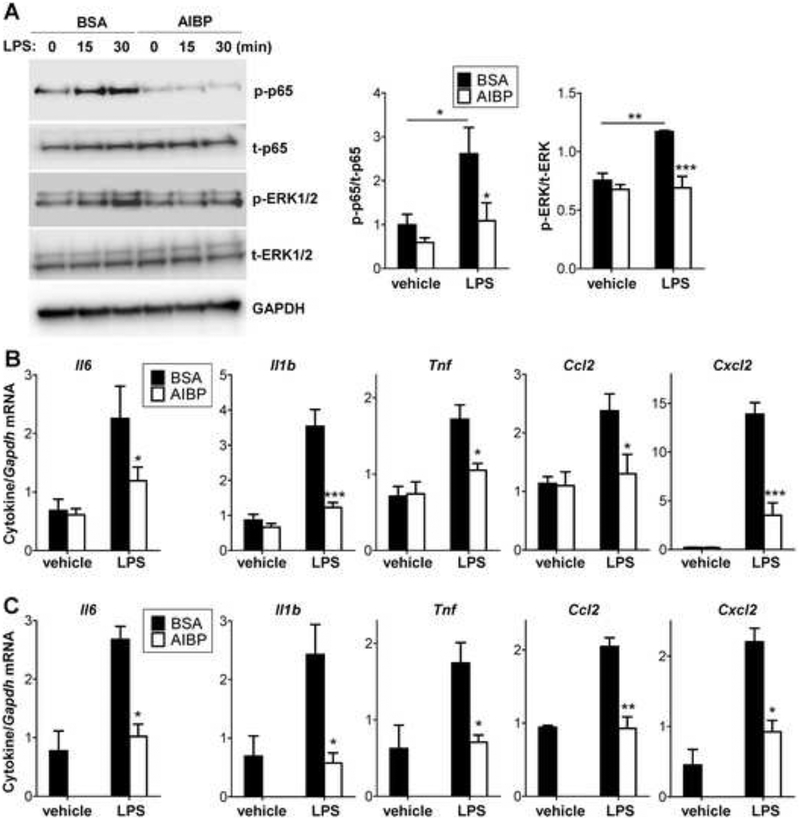
Figure 3. AIBP reduces inflammatory responses in microglia.
A-B, BV-2 cells were incubated for 2 hours with 0.2 μg/ml BSA or AIBP in serum-containing medium and stimulated with 10 ng/ml LPS. p65 and ERK1/2 phosphorylation were tested after 30 min (A), and cytokine mRNA expression after 2 h of incubation (B). C, Primary mouse microglia (pooled from 5–6 mice per sample) were incubated for 2 hours with 0.2 μg/ml BSA or AIBP in serum-containing medium and stimulated with 10 ng/ml LPS for 1 hour. Mean±SEM; n=4–6 for BV-2; n=3 for primary microglia experiments; *, p<0.05; **, p<0.01; ****, p<0.0005 (Student’s t-test). Due to limited availability of primary cells, ‘vehicle/AIBP’ group was omitted. See also Figures S3 and S4A.

