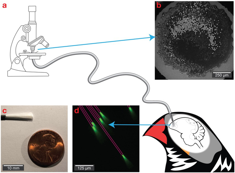
Figure 1. Bundles of microfibers as a potential deep brain optical interface.
(a) The polished imaging surface is mounted in a traditional fluorescence microscope, while individual fibers with a diameter as small as 6.8 μm are implanted into the brain. (b) The polished imaging surface that connects with the microscope. (c) A bundle of 18,000 fibers. (d) Light propagates with near total internal reflection, allowing it to deliver and collect light at the tips of the fibers. Six fibers are shown in a fluorescein solution, with pink lines added to emphasize fiber path.

