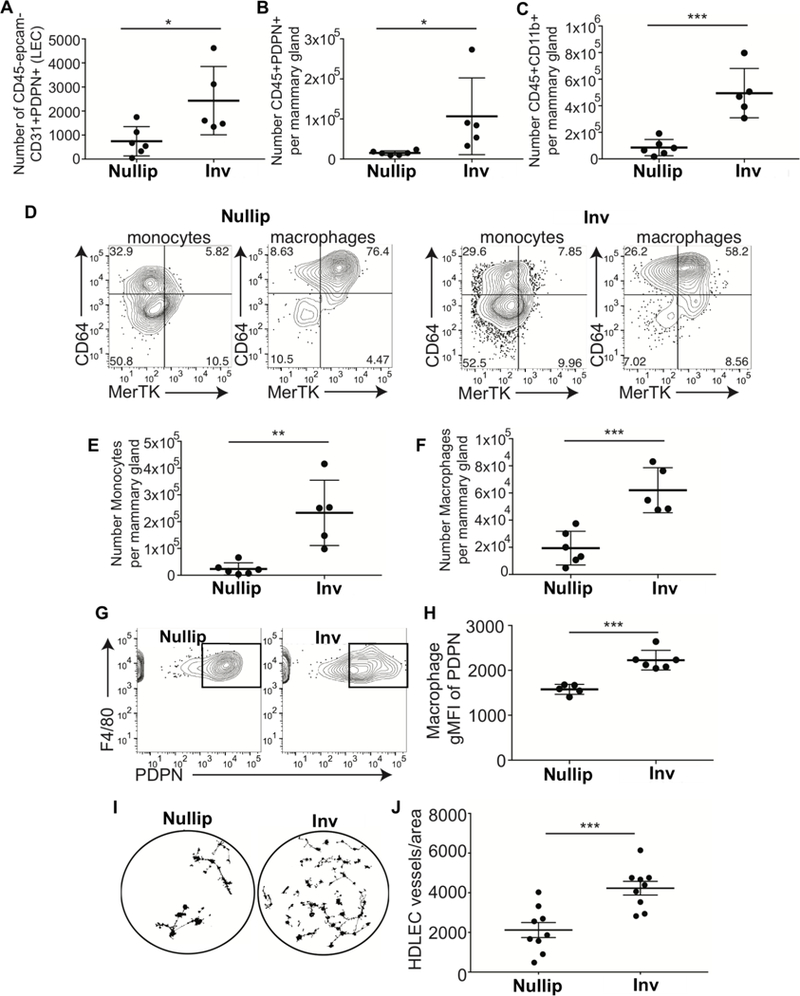
Figure 2: Podoplanin expression on macrophages drives lymphangiogenesis.

Flow cytometry analysis of cells isolated from nulliparous or involution day 6 mouse mammary tissues reveals A) increased lymphatic endothelial cells, B) PDPN+ leukocytes, C) PDPN+ myeloid cells, D-F) monocytes and macrophages, as well as G&H) PDPN-expressing F4/80+ macrophages. I&J) F4/80+ macrophages isolated from nulliparous and involution day 6 mouse mammary glands enhance in vitro lymphangiogenesis. Unpaired t-test: *p<0.05, **p<0.01, ***p<0.001.
