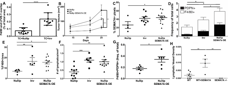
Figure 3: Semaphorin 7a promotes tumor-associated macrophages and lymphangiogenesis.

A) In an in vivo matrigel plug assay, 66cl4 tumor cells were mixed with macrophages isolated from nulliparous or recently weaned mice (at involution day 6). The resulting plugs were harvested and F4/80/LYVE-1 co-staining quantitated. B) Growth of 66cl4 cells engineered to overexpress SEMA7A compared to empty vector controls injected into nulliparous and involution day 1 mouse mammary tissues. C) SEMA7A expression by IHC in tumors from B. D) CD45+CD31-F4/80+PDPN+ cells in tumors from B. E) %F4/80+ cells and F) # LYVE-1+ vessel density in tumors from B. G) F4/80+PDPN+ cells in E0771 SEMA7A tumors in nulliparous mice. H) LYVE-1+ density in tumors from G with the addition of tumors from SEMA7A−/− hosts injected with E0771 cells. Unpaired t-test: *p<0.05, **p<0.01, ***p<0.001.
