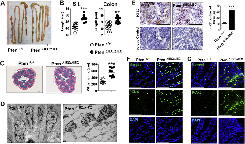
Figure 2. Intestinal epithelial cells of Pten ΔIEC/ΔIEC mice exhibited enhanced mitotic activity compared to those of Pten +/+ mice.

(A and B) Presented is the representative gross appearance of the intestine from Pten ΔIEC/ΔIEC and littermate Pten +/+ mice (A). Full length of the small intestine and the colon from age (1 year)- and sex- matched mice was evaluated (n=14/group) (B). (C) Paraffin-embedded sections of the mid-colon were prepared from age (3 months)-and sex-matched Pten ΔIEC/ΔIEC (n=9) and littermate Pten +/+ (n=9) mice. The sections were subjected to Alcian blue staining, followed by measurement of villus height under Axio Imager Z1 microscope (Carl Zeiss, Oberkochen, Germany). (D) Presented are electron micrographs of the colonic epithelium from Pten ΔIEC/ΔIEC and littermate Pten +/+ mice at the age of 12 month. (E) With the frozen sections of the mouse colon, expression of the cell proliferation marker Ki-67 was evaluated by immunohistochemistry with an antibody against Ki-67 and its isotype control IgG. Ki-67 positive cell index was evaluated and presented as means ± SEM. (F and G) Paraffin-embedded sections of the mid-colon were subjected to immunofluorescence staining with PCNA (F) and phospho-Akt (P-Akt) (G) antibodies. ** P<0.01, *** P<0.001 (Mann-Whitney U test). Representative images from three independent experiments were presented. Each bar in the graph indicates mean. Scale bar represents 100 μm (E) or 50 μm (F, G).
