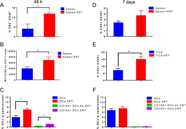
Figure 5.

XRT induced the expression of OX40 and upregulated antigen-presenting dendritic cells. A, B, C, anti-PD1 resistant tumor-bearing mice were treated with XRT 12 Gy x 3 (n=3-5 mice per group) and splencoytes were analyzed by flow cytometry 48 h after the last dose of XRT. A, Percentages of CD4+ OX40+ T-cells B, mean fluorescence intensity (MFI) values of OX40 expression on CD4+ T-cells. C, Percentages of Dendritic cells (DCs) and CD103+ subset of DCs were reported. D, E, F, splenocytes and tumor-infiltrating lymphocytes (TILs) were analyzed by flow cytometry 7 days after XRT as in A, B, C. For OX40 expression, cells were first gated on CD45+, then on CD3+, followed by CD4+ T-cells. To analyze the dendritic cell percentages, cells were gated on CD45+, then CD3−CD11c+, or CD3−CD11c+ CD103+. *P ≤ 0.05 is considered significant.
