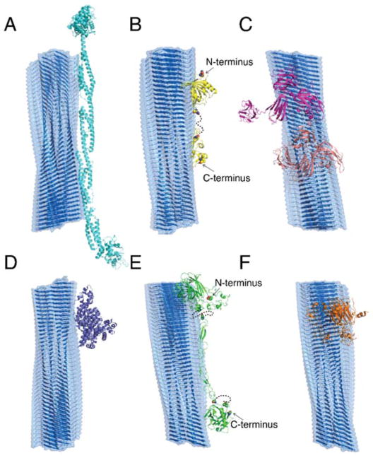
Figure 5. Schematic binding between IAPP amyloid fibril and AF/AE proteins with maximal contact interface.
The amyloid fibril is formed by β-sheets of composite peptides (grey). The binding of AF proteins – (A) alpha-actinin-4 (cyan), (B) protein AMBP (yellow) and (C) neuropilin in open (magenta) and closed (light pink) conformations – and AE proteins – (D) serum albumin (purple), (E) thrombospondin-1 (blue) and (F) cartilage oligomeric matrix protein (orange) – with the amyloid fibril was estimated by aligning them with maximum contact surface areas. Serum proteins are shown in cartoon format with dashed lines representing missing sequences without available structural information.

