
Fig. 7. FGF4 and TGFβ2/TGF0β3 induce chondrogenesis.
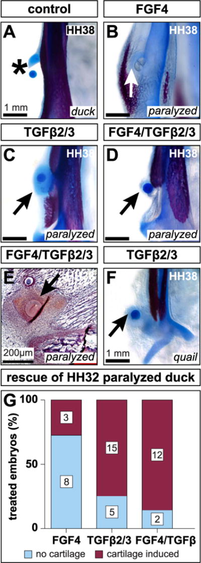
(A) Ventral view of a cleared and stained mandible treated with a BSA soaked bead. Carrier treatments exert no effect on secondary cartilage (asterisk). (B) HH32 FGF4 treatment induces cartilage (arrow) in paralyzed embryos by HH38. (C) TGFβ2/TGFβ3 treatment induces cartilage (arrow) in paralyzed embryos. (D) Combined FGF4 and TGFβ2/TGFβ3 treatments induce cartilage (arrow) despite paralysis. (E) HH38 sagittal section through the mandibular adductor insertion of a paralyzed embryo implanted with FGF4 and TGFβ2/TGFβ3 beads at HH32. Safranin-O reveals dense, positively stained mesenchyme surrounding the beads (arrow). (F) HH32 TGFβ2/TGFβ3 treatment induces quail to form cartilage by HH38 (arrow). (G) FGF4, TGFβ2/TGFβ3, and FGF4/TGFβ2/TGFβ3 treatments induce cartilage by HH38. The distribution of treatment outcomes depends upon the ligand or ligands embryos receive (Fisher’s Exact Test p = 0.005).