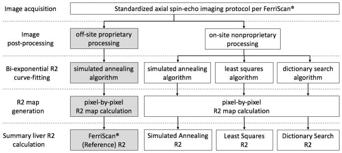
Figure 1.
Flowchart describes the R2-MRI relaxometry procedures step-by-step from image acquisition to summary liver R2 calculation. The proprietary procedures performed off-site by Resonance Health are indicated by gray boxes. Open boxes represent on-site procedures. The three nonproprietary methods differ in the curve-fitting algorithm but share an identical image post-processing step. The on-site image processing was independently implemented following the general principles of those used in the proprietary method.

