
Figure 7. Canonical ultrastructural features of Mcoln-/- mice compared to aged heterozygous and WT mice.
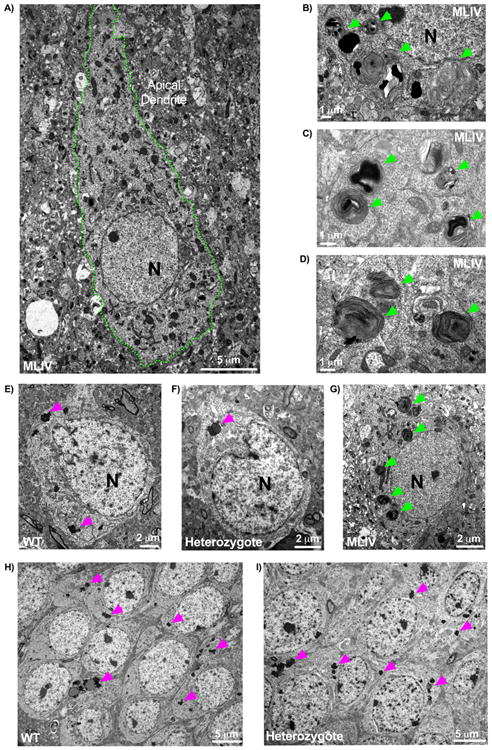
A) Characteristic pathology in Mcoln1-/- 3-month old tissues revealed by electron microscopy. Shown here is a neocortical neuron with the neuronal cytoplasm found congested with multiple compound, electron dense storage bodes as well as granulomembranous storage bodies and an enlarged apical dendrite (cell membrane outlined in green). B-C) Electron dense, compound storage bodies typically found in 3-month-old mutant mice, taken here from neocortex and highlighted by green arrows. E-G) Neocortical neurons from (E) WT, (F) heterozygote, and (G) mutant. E-F) WT and heterozygote neocortex exhibited neurons possessing lipofuscin accumulation (pink arrowheads) but otherwise devoid of pathological, compound storage bodies readily detectable in the mutant (G, green arrowheads). Neither heterozygous or WT neurons (both shown here at 18+ months) were found to exhibit abnormal structures, such as the swollen apical dendrites captured in 3-month old mutant neocortex (A). H-I) Hippocampal sections from WT (H) and heterozygous (I) mice. Similar to findings in cerebral cortex, hippocampal neurons displayed comparable features between WT (H) and heterozygotes (I) such as lipofuscin granulation storage with no evidence of pathological storage. Lipofuscin was not found to be quantitatively increased in the heterozygote in either cerebral cortex or hippocampus (data not shown). All tissues shown here are representative images from mice aged 18+ months, with the exception of the mutant at 3 months. 2 heterozygotes, 4 WT, and 4 Mcoln1-/- mice were probed for pilot EM data.