Fig. 1.
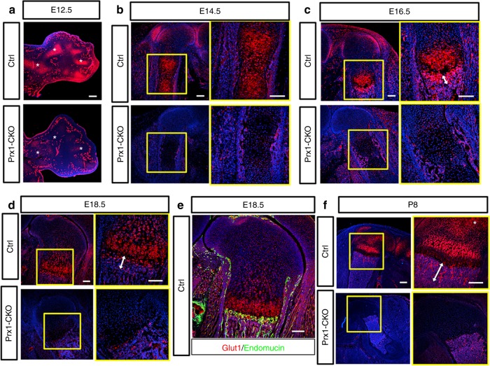
Genetic deletion with Prx1-Cre eliminates Glut1 in the long bones of the mouse. a Immunofluorescence staining for Glut1 on longitudinal sections of the forelimbs of E12.5 control (Ctrl) or mutant littermates (Prx1-CKO). Asterisk denotes cartilage elements. Note comparable expression in the vasculature between control and mutant embryos. b–f Glut1 immunostaining on longitudinal sections of the femur of littermate control or mutant embryos at E14.5 (b), E16.5 (c), E18.5 (d, e), and P8 (f). Boxed areas are shown at higher magnification to the right. Endomucin immunostaining in green marks endothelial cells (e). Double arrowheads denote trabecular bone region (c, d, f). Scale bar: 100 µm (a–e) or 200 µm (f). Each image is representative of three animals

