Fig. 3.
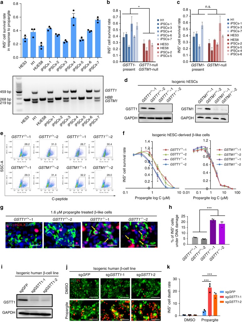
A hPSC-based population study discovers that GSTT1-null pancreatic β-like cells are hypersensitive to propargite-induced cell death. a Survival rate of INS+ cells derived from 10 different hESC or iPSC lines cultured in the presence of 1.6 μM propargite (n = 3), and genotype analysis of GSTM1 and GSTT1 in those hESCs and iPSCs. b, c Correlation of INS+ cell survival rate in the presence of 1.6 μM propargite in cells lacking both GSTM1 and GSTT1 (b), or lacking only GSTM1 (c). n.s. indicates a non-significant difference. d Western blotting analysis of GSTT1 or GSTM1 protein expression in INS+ cells derived from isogenic wild type, GSTT1−/− or GSTM1−/− H1 hESCs. The −/− null clones were CRSIPR-induced biallelic frameshift mutants. The two GSTT1 knockout clones were both homozygous null mutants, and the two GSTM1 knockout clones were both compound-null mutants. e Flow cytometry analysis of C-peptide+ cells in isogenic GSTT1−/− or GSTM1−/− hESC-derived D18 cells. f Inhibition curve of propargite on INS+ cells derived from GSTT1+/+ or GSTT1−/− H1 hESCs (n = 3). g, h Representative images (g) and DNA damage rate (h) of GSTT1+/+ and GSTT1−/− β-like cells (n = 3). Scale bars, 800 μm. γ-H2A.X +/INS+ cells are highlighted with yellow arrows. i Western blot analysis of GSTT1 protein in EndoC-βH1 cells carrying sgGSTT1. Two CRISPR gRNAs (sgGSTT1-1 and sgGSTT1-2) were used for generating GSTT1−/− EndoC-βH1 cells. j, k Representative images (j) and cell death rate (k) of GSTT1−/− EndoC-βH1 cells treated with 1.6 μM propargite (n = 3). Scale bars, 200 μm. Values presented as mean ± S.D. n.s. indicates a non-significant difference. p values calculated by unpaired two-tailed Student’s t-test were *p < 0.05, ***p < 0.001. Related to Supplementary Fig. 3

