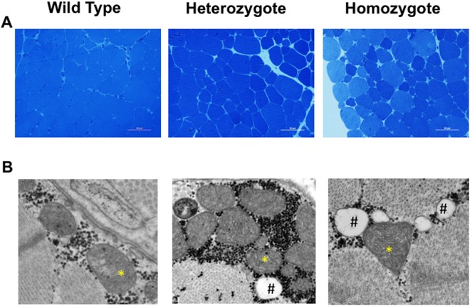
Figure 5.
Skeletal muscle histological phenotype in ALPL c.1077 C > G targeted sheep. (A) Representative light microscopic evaluation (40X magnification, size bar, 50 µm) of Richardson’s stained 500 nm sections of 2 month old gluteus muscle reveal variable sized muscle fibers in mutants compared to homogeneously sized WT fibers. (B) Representative electron micrographs (original magnification 44,000X) of HPP mutants compared to WT reveal abnormal mitochondria cristae ultrastructure (*) and higher fat content (#).

