Figure 1.

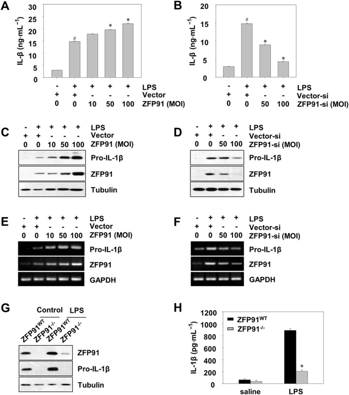
ZFP91 up‐regulates the production and activation of pro‐IL‐1β in LPS‐stimulated THP‐1 cells and BMDMs. (A, C) PMA‐differentiated THP‐1 cells were transduced with lentiviruses containing ZFP91 gene using multiplicities of infection (MOI) of 10, 50 and 100 for 24 h and then stimulated with 1 μg·mL−1 LPS for 6 h. IL‐1β was detected by ELISA. Data are the mean ± SD of five independent experiments. # P < 0.05 versus control group (cultured in medium alone); *P < 0.05 versus LPS‐induced group. The protein levels of pro‐IL‐1β were measured by Western blot analysis. (B, D) PMA‐differentiated THP‐1 cells were transduced with lentiviruses carrying siRNA against ZFP91 using MOI of 50 and 100 for 24 h and then stimulated with 1 μg·mL−1 LPS for 6 h. IL‐1β was detected by ELISA. Data are the mean ± SD of five independent experiments. # P < 0.05 versus control group (cultured in medium alone); *P < 0.05 versus LPS‐induced group. The protein levels of pro‐IL‐1β were measured by Western blot analysis. (E) PMA‐differentiated THP‐1 cells were transduced with lentiviruses containing ZFP91 gene using MOI of 10, 50 and 100 for 24 h and then stimulated with 1 μg·mL−1 LPS for 6 h. The mRNA level of pro‐IL‐1β was measured by RT‐PCR analysis. (F) PMA‐differentiated THP‐1 cells were transduced with lentiviruses carrying siRNA against ZFP91 using MOI of 50 and 100 for 24 h and then stimulated with 1 μg·mL−1 LPS for 6 h. The mRNA level of pro‐IL‐1β was measured by RT‐PCR analysis. (G) BMDMs from wild type (WT) or ZFP91 knockout (ZFP91−/−) C57BL/6 mice stimulated with 1 μg·mL−1 LPS for 6 h. The protein levels of pro‐IL‐1β were measured by Western blot analysis. (H) BMDMs from WT or ZFP91 knockout (ZFP91−/−) C57BL/6 mice stimulated with 1 μg·mL−1 LPS for 6 h. IL‐1β was detected by ELISA. Data are the mean ± SD of five independent experiments. *P < 0.05 compared with LPS‐treated WT mice.
