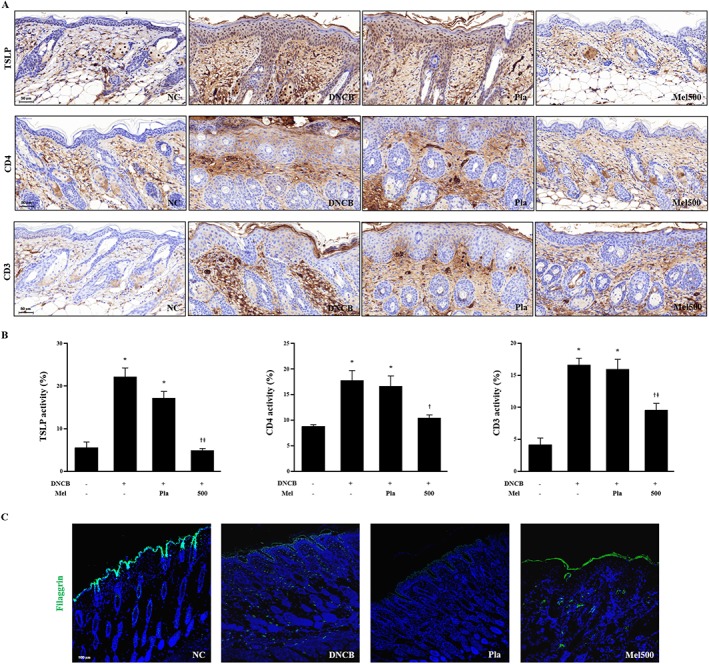
Figure 5.

Effects of melittin on TSLP expression, CD4+ and CD3+ T cells in DNCB‐sensitized mice. (A) Histological images and (B) graphs indicating the relative percentage of TSLP expression and CD4+ and CD3+ immunopositive cells of the dorsal skin sections (n = 5). (C) Effects of melittin on abnormal epidermal differentiation. Immunofluorescence staining of sections of skin with antibody specific for filaggrin shows the expression of epidermal differentiation markers as labelled with FITC, green. Cells were counterstained with Hoechst 33342 (blue). Scale bar = 100 μm. *P < 0.05 versus NC; + P < 0.05 versus DNCB; ǂ P < 0.05 versus Pla; NC, normal control; DNCB, DNCB‐sensitized and challenged; Pla, placebo; Mel500, melittin 500 μg.
