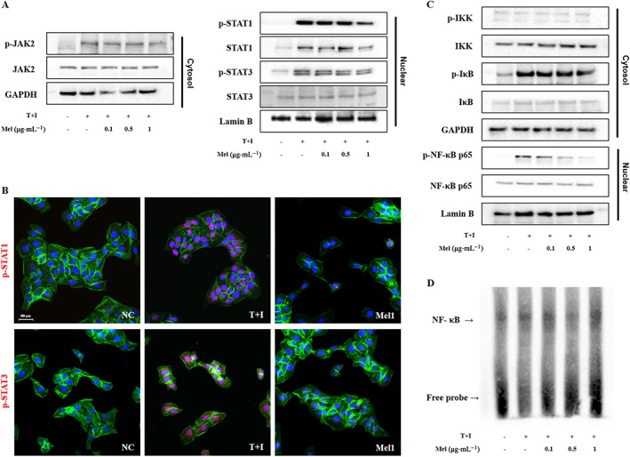
Figure 7.

Effects of melittin on activation of the (A) JAK2, STAT1 and STAT3 signalling pathways in TNF‐α/IFN‐γ‐stimulated HaCaT cells. (B) Immunofluorescence staining for p‐STAT1 and p‐STAT3 (labelled with Alexa Fluor 555, red), and F‐actin (labelled with Alexa Fluor 488, green). Cells were counterstained with Hoechst 33342 (blue). Effects of melittin on activation of the (C) NF‐κB signalling pathway in TNF‐α/IFN‐γ‐stimulated HaCaT cells. (D) NF‐κB DNA binding activity in the nuclear extract was measured by EMSA. Representative images from each group. Scale bar = 50 μm. NC, normal control; T+I, TNF‐α/IFN‐γ‐stimulated; Mel1, TNF‐α/IFN‐γ‐stimulated +1 μg·mL−1 Mel.
