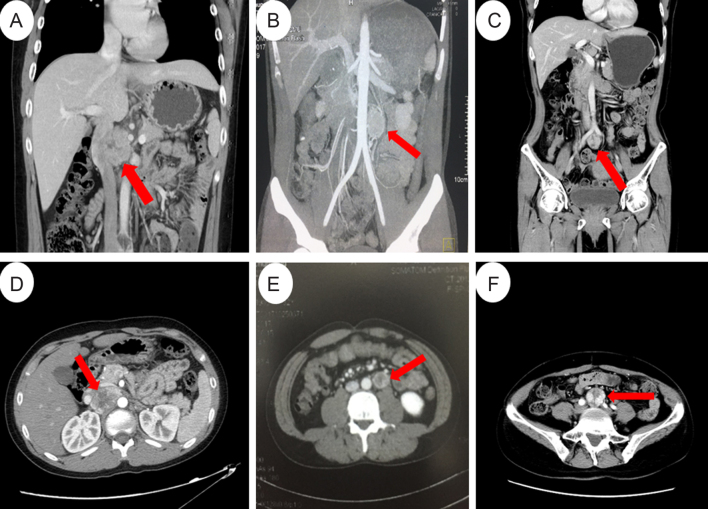
Figure 2.
Abdominal CT or MRI scans of the probands. (A and D) Coronal (A) and axial (D) CT images of the 5.1 × 3.4 cm retroperitoneal mass between the aorta and inferior vena cava in proband 1. (B and E) Coronal (B) and axial MRI (E) images of the 3 × 2 × 2 cm retroperitoneal para-aorta mass in proband 2. (C and F) Coronal (C) and axial (F) images of the 2.9 × 2.7 cm mass located at the bifurcation of the abdominal aorta in proband 3.

 This work is licensed under a