Fig. 1.
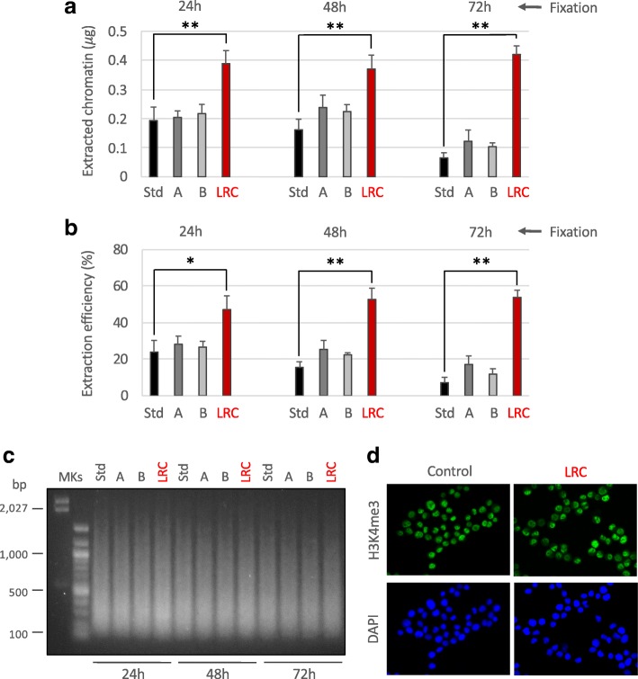
Attempts to improve chromatin extraction from FFPE samples at different times of fixation. Different conditions of chromatin extraction from normal colon FFPE tissues fixed at the times reported in figure were tested. The total amount of isolated chromatin was fluorimetrically evaluated after chromatin de-crosslinking and DNA purification (a) while the extraction efficiency was calculated considering the amount of DNA of extracted chromatin compared to the total DNA present in the sample (b). Std: standard PAT-ChIP, 18 pulses of sonication of 5 s at 85% of amplitude; A: 54 pulses of sonication of 5 s at 75% of amplitude; B: 54 pulses of sonication of 5 s at 65% of amplitude; LRC: condition in which the sample was subjected to limited reversal of crosslinking, 3 pulses of sonication of 30 s at 40% of amplitude. *P < 0.05 with respect to standard condition for each time of fixation by one-way ANOVA with Tukey’s HSD. **P < 0.01 with respect to standard condition for each time of fixation by one-way ANOVA with Tukey’s HSD. All the experiments were conducted in triplicates. c Evaluation of chromatin fragmentation by electrophoretic separation on 1.3% agarose gel electrophoresis (AGE) followed by SYBR Gold staining of purified input DNA. MKs, molecular weight markers. d Compatibility of LRC with H3K4me3 immunoselection. HeLa cells were subjected to formaldehyde fixation and treated with LRC or left untreated. Cells were stained by immunofluorescence with anti-H3K4me3 antibody (green, upper panels) following the same procedure described for the PAT-ChIP assay (buffers, timing, and temperature of incubations) and with DAPI to label nuclei (blue, lower panels)

