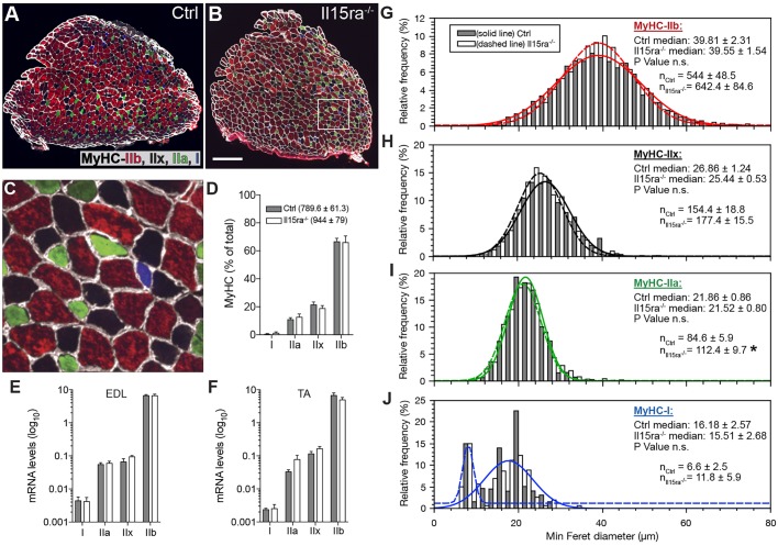
Fig. 2.
Effect of lack of IL15RA on muscle fiber type. (A–C) MyHC staining in control (A) and Il15ra−/− (B) EDL muscles. The area enclosed in the white box is shown at 6× magnification in C. Scale bar: 200 µm (D) Relative fiber-type composition of EDL (n=5, counting all the fibers in each section). The mean±s.e.m. number of fibers per section is shown in the legend. Quantifications of MyHC isoforms by qPCR in EDL (E) and TA (F) samples (n=4). (G–J) Frequency histograms of the quantification of minimum Feret diameter of EDL fibers distinguishing between IIb (G), IIx (H), IIa (I), I (J) fibers (n=5 mice per genotype, measuring all the fibers in each section, *P<0.05).

