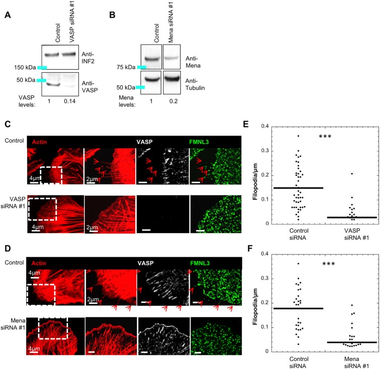
Fig. 4.
Suppression of VASP or Mena causes filopodial reduction in U2OS cells. (A) Western blot of U2OS cells treated with control siRNA or VASP siRNA#1, then probed for VASP or INF2 (loading control). Numbers beneath lanes indicate the relative amount of VASP remaining. (B) Western blot of U2OS cells treated with control siRNA or Mena-siRNA #1, then probed for Mena or tubulin (loading control). Numbers beneath western lanes indicate the relative amount of of Mena remaining. All lanes are from one gel, with intervening lanes removed. Image processing of all lanes is identical. (C,D) U2OS cells treated with control siRNA, VASP siRNA #1 or Mena siRNA #1 were fixed and stained with TRITC–phalloidin (red), and anti-FMNL3 and anti-VASP antibodies. Micrographs are maximum intensity projections (MIPs) of 0.18 μm z-slices (4–8 slices); red arrows indicate filopodia. (E,F) Dot plots of filopodial density for VASP and Mena knockdown, respectively. (E) Control (mean 0.152±0.086) and VASP siRNA #1 (mean 0.028±0.043) n=30 cells, five experiments. (F) Control (mean 0.171±0.079) and Mena siRNA #1 (mean 0.039±0.051) n=32 cells, three experiments. ***P-value <0.0001 as calculated by Mann–Whitney U-test. Errors are given as s.d.

