
Fig. 2.
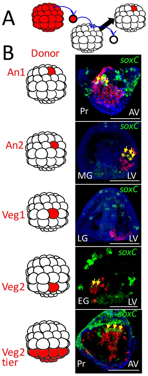
Cell lineage analysis indicates that neurogenesis is initiated in three locations in the embryo. (A) Schematic of the experimental procedure. At the 60-cell stage, a single RFP-expressing cell (red) is transplanted to an equivalent location in an unlabeled 60-cell stage embryo. The donor is from the same tier of cells as the cell it replaces as shown in the diagrams to the left of the images in B (the top four tiers are named An1, An2, Veg1 and Veg2 as indicated (the bottom two tiers, the large and small micromeres, have known fates and so were not considered). (B) Embryos were fixed at different stages and double in situ hybridization performed to identify soxC expression (green), which by this time exclusively marks neural progenitors. The patch of cells derived from the RFP donor is stained red. The yellow arrows point to soxC-positive cells that also are stained red, thus neural lineage progeny of the labeled donor cell. Of more than 80 cases scored we observed no double-labeled neural progenitors outside the patch of donors, including the bottom image in which the entire Veg2 tier was transplanted. In that image, as expected, a number of cells migrate from the patch but none of the migrants is both red and green. In each case, a portion of a confocal stack is shown at the level of double-labeled cells, if present. Transplant cases scored: An1, n=3; An2, n=18; Veg1, n=25; Veg2, n=30; Veg2 tier, n=4. AV, animal view; EG, early gastrula; LG, late gastrula; LV, lateral view; MG, midgastrula; Pr, prism. Scale bars: 50 µm.