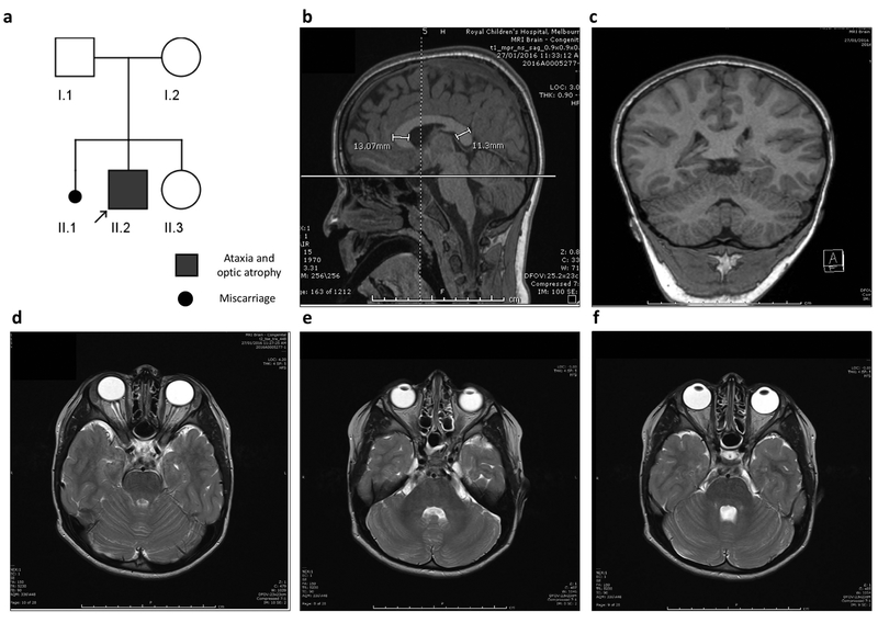
Figure 1. Pedigree of SLC25A46 patient and MRI images.
(a) Pedigree of index patient (arrow) homozygous for the c.770G>A p.Arg257Gln (p.R257Q) variant. Both parents were confirmed carriers for the mutation. (b-f) Sagittal, coronal, and serial axial MRI images obtain at the age of 5½ years, demonstrating normal appearance of the cerebellar vermis and hemispheres. The corpus callosum is diffusely thick with the genu slightly thicker than the splenium (b). The cerebellum and optic nerve appearance are within the normal limits for age.

