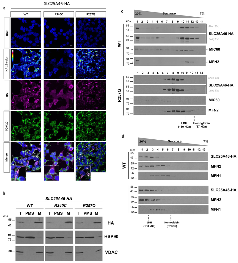
Figure 4. Analysis of SLC25A46 expression and native molecular weight in stable transfected HEK293T cells.
(a) Immunocytochemistry of cells expressing wildtype (WT), p.R257Q, or p.R340C variants. DAPI was used to visualize nuclei and an anti-TOM20 antibody was used as mitochondrial marker (b) Cell fractionation analysis of wildtype (WT), p.R257Q, and p.R340C transfected cells. Total cell lysate (T), post-mitochondrial supernatant (PMS), representing the cytosolic soluble fraction, and isolated mitochondria (M) were analyzed by SDS-PAGE and immunostaining with antibodies against HA tag, HSP90, as cytosolic marker, and VDAC, as mitochondrial marker. (c) Sedimentation of wild-type SLC25A46-HA and p.R257Q in a linear 7–20% sucrose gradient centrifuged in a Beckman 55Ti rotor at 28,000 rpm for 12 hours. The proteins hemoglobin (67 kDa) and LDH (130 kDa) were used to calibrate the gradient. (d) Same as (c) although gradients were centrifuged at 45,000 rpm for 13 hours to resolve the lighter MFN2-containing complexes.

