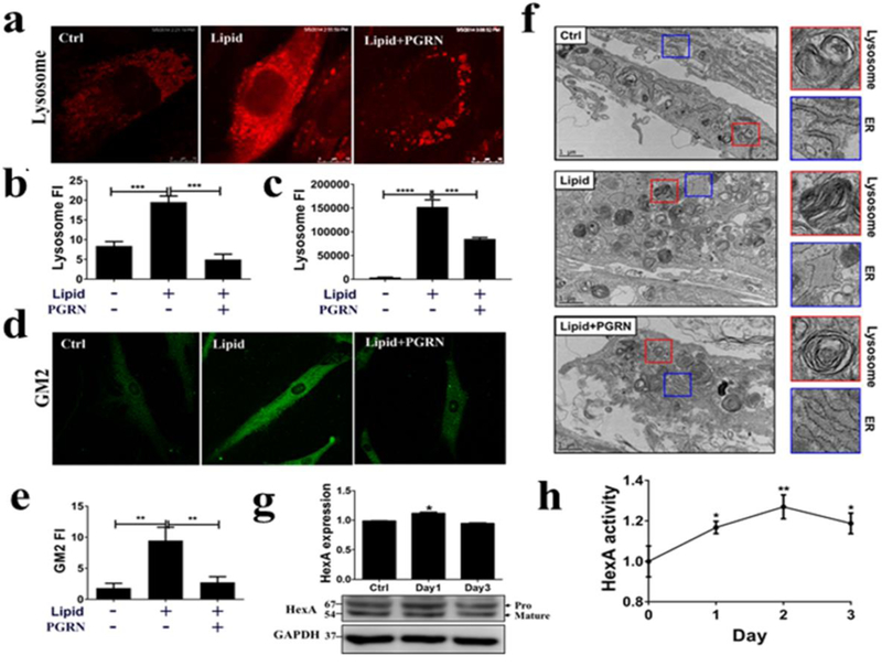
Fig. 4. Therapeutic effect of PGRN acts through increased activity of HexA.

Fibroblasts from TSD patients were treated with PBS, lipid (50 μg/ml) and PGRN (0.5 μg/ml) for 24 hours. (a) The staining of lysosomes by LysoTracker Red (300 nM). PGRN significantly reduced lysosomal storage. (b) The quantification of the red fluorescence of (a). (c) After LysoTracker Red staining, lysosomal signals were detected by the plate reader, SpectraMax ® i3. (d) GM2 staining by immunofluorescence. PGRN greatly reduced GM2 accumulation. (e) The quantification of the GM2 staining shown in (d). (f) Transmission electronic microscope analysis of morphological changes in lysosomes and ER following lipid challenge and PGRN treatment. Fibroblasts were challenged with lipid lysate, or lipid plus recombinant PGRN proteins (0.5 μg/ml) for 3 days. The cells were processed for TEM analysis. The morphological changes of lysosomes (red square) and ER (blue square) were observed following treatment of PGRN. (g) PGRN (0.5 μg/ml) increased HexA expression level in TSD fibroblasts. TSD fibroblasts were treated with PGRN for 3 days. The HexA protein levels were examined by western-blot. The figure is representative of three independent experiments. (h) PGRN enhanced HexA enzymatic activity in TSD fibroblasts. TSD fibroblasts were treated with PGRN (0.5 μg/ml) for 3 days. The enzymatic activities were standardized by control group and measured by processing its specific substrate, MUGS. Data are presented as mean ± SEM. *P<0.05. **P<0.01. ***P<0.001. ****P<0.0001.
