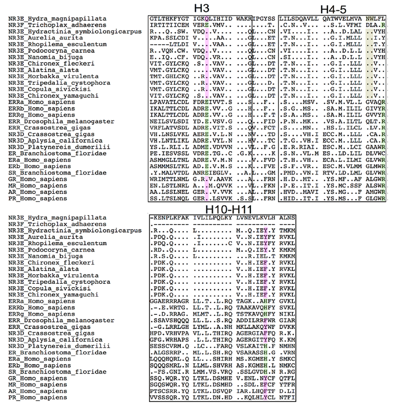
Figure 5. Alignment of H3, H4-H5 and H10-H11 helices in the NR3 family, compared to NR3E from Hydra.

Residues involved in estrogen binding in ER are highlighted in green. Homologous residues that are shared by the cnidarian NR3E and the vertebrate oxosteroid receptors (SRs) are highlighted in pink. The cnidarian-specific N79-W80 anchor is highlighted in brown. An alignment for the entire DNA-binding domain and ligand-binding domain is shown in Supplemental Figure 2. The helices from the ligand-binding domain are mapped according to the structure of human ERα [48], Calculations of identity percentages for both domains are also provided in Supplemental Table1.
