
Figure 6. A possible pathway for steroid enterocrine signaling in cnidarians.
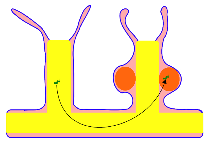
Steroids, in green, could be produced from dietary sterols in the gut (in yellow) and go to the gonad (in orange) along with other nutrients. Gonads may be of ectodermal or endodermal origin, depending on the species. The pink tissue represents the mesoglea, which consists of a gelatinous matrix that contains collagen fibers and usually some cells. The mesoglea forms a hydrostatic skeleton, but does not contain a circulating body fluid or play a known role in circulation.