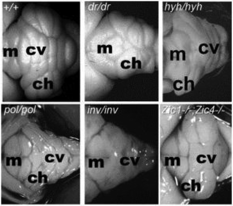
Figure 2. Examples of mouse cerebellar malformations.
Dorsal whole mount views of cerebellar malformations in 4 spontaneous mutants and 1 engineered mouse strain. Wild-type (+/+) cerebellum with cerebellar vermis (cv) and cerebellar hemispheres (ch) indicated showing stereotypical foliation pattern. Disruption of this patterning is obvious in many mouse mutant strains. For example, in dreher (dr) homygous mutants, a reduced cerebellar vermis causes juxtaposition of the cerebellar hemispheres. In hydrocephalus with hop gait (hyh) homozygous mutants, the vermis is more prominent than the hemispheres. Although these mice are not models for any specific human malformation, investigation of the underlying pathogenesis has provided insights into the role of the roof plate in cerebellar development and vermis formation. Polaris (pol) and inversus (inv) homozygous mutants have severely disrupted cerebellar morphology and are models for cilia related MTM human cerebellar malformations. Zic1/4 double homozygous mouse mutants model human DWM and display simplified vermis foliation. Anterior is to the left, indicated by the presence of midbrain colliculi (m). Photos are optimized to show pattering differences and are not all at the same magnification.

