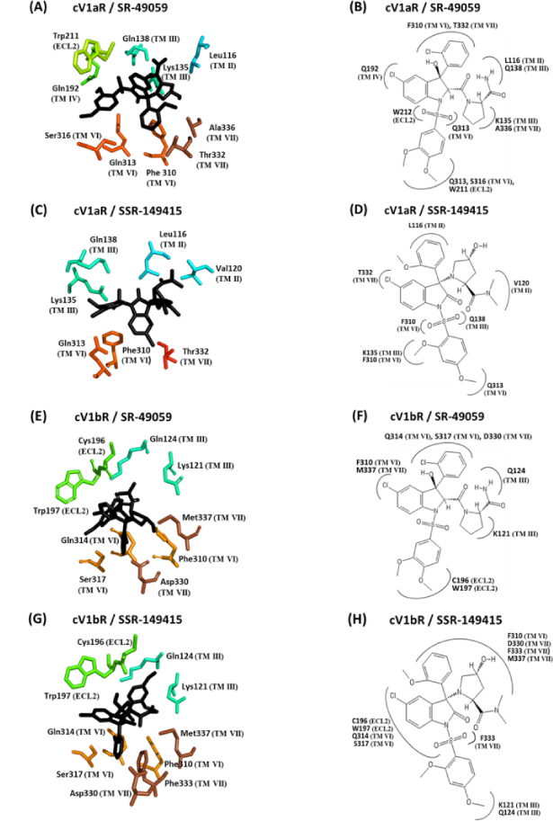
Figure 2. Close-up and two-dimensional (2D) schematic views of the interacting amino acid residues of cV1aR and cV1bR with SR-49059 and SSR-149415.

(A), (C), (E), and (G); Antagonists are shown as sticks in black color and the amino acid residues involved in the interaction are shown in different colors according to the transmembrane helices (TM) or extracellular loop (ECL). The residues are numbered according to their position in the primary sequence (see Fig 3). The seven TM helices are shown in different colors. (B), (D), (F), and (H); 2D schematic view of the interaction models with antagonists. All of the amino acid residues potentially interacting with the different parts of the antagonists are shown. Numbering of the residues and of the TM is equivalent to that used in Fig 3.
