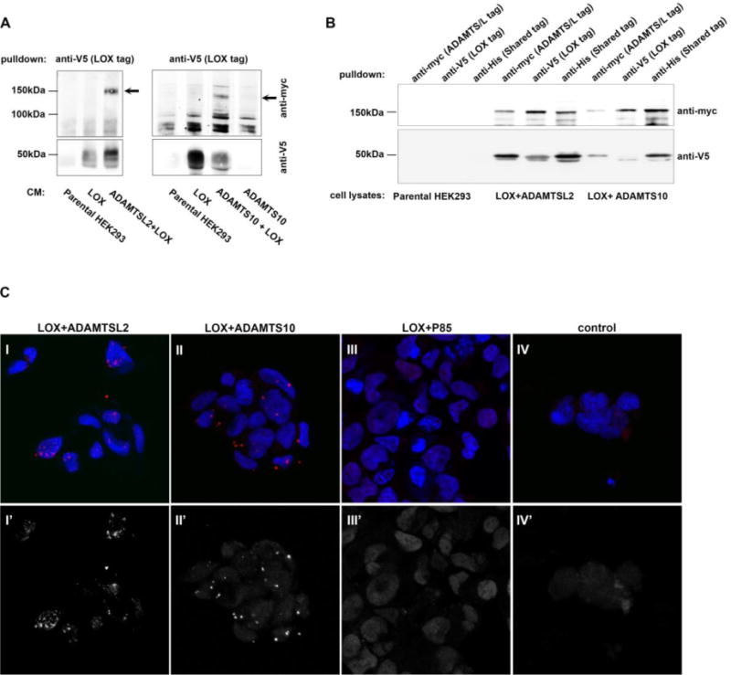
Figure 2. LOX forms a complex with ADAMTSL2 and ADAMTS10.

A. Co-immunoprecipitation of 4 day conditioned medium from stably transfected HEK293 cells expressing LOX plus ADAMTSL2, ADAMTSL2 alone (negative control), LOX plus ADAMTS10, or ADAMTS10 alone (negative control). B. Co-immunoprecipitation of stably transfected HEK293 cells expressing LOX plus ADAMTSL2 or LOX plus ADAMTS10 or parental HEK293 lysate (negative control). LOX migrates as two bands at ~50 kDa. ADAMTSL2 at ~150 kDa, and ADAMTS10 at 150 kDa. Membranes (A,B) were incubated with anti-myc (top) and anti-V5 (bottom). C. Proximity ligation assays performed on HEK293 cells expressing LOX+ADAMTSL2 (I, I′ IV, IV′), LOX+ADAMTS10 (II, II′) and LOX+P85 (III, III′). Anti-LOX and anti-myc antibodies were used to target LOX and ADAMTSL2/ADAMTS10 or P85, respectively. Signal amplification, marked by red signal (top, I-IV) or shown in grayscale (bottom, I′–IV′) is observed only in cells expressing LOX and an ADAMTS protein. In panel IV (control), no primary antibodies we added.
