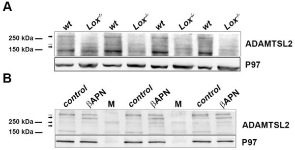
Figure 6. Alterations in putative ADAMTSL2 complexes following Lox deletion or inhibition.

Upper parts of the gels shown in Figs. 4E and 5. A. ADAMTSL2 western blot from E18.5 wild-type and Lox−/− brain lysates. B. Western blot of ADAMTSL2 in adult control and βAPN-treated mouse aorta lysates. Arrows mark high molecular weight bands which are affected (black) or not affected (grey), by the loss of Lox (A) or βAPN treatment (B). Membranes were incubated with anti-ADAMTSL2. M designates molecular weight marker.
