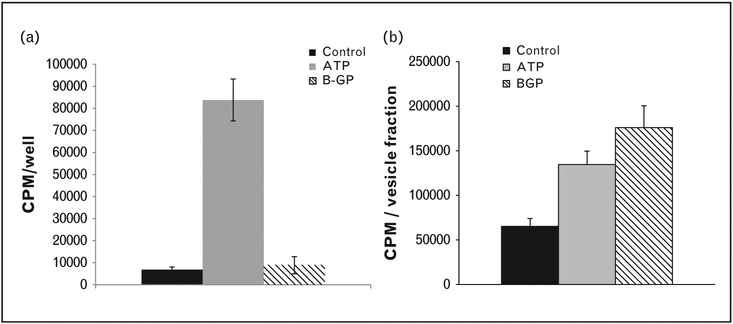
FIGURE 3.
Mineralization patterns differ in chondrocyte monolayers and ACVs in response to ATP and β-glycerophosphate. (a) Adult porcine chondrocyte monolayers were incubated with 1mM ATP or 1mM β-glycerophosphate (BGP) in media trace labeled with 45Ca2+. After 48 h, media were removed and 45Ca2+ in the cell layer was quantified with liquid scintigraphy. (b) ACVs isolated from adult porcine cartilage were incubated in a solution trace labeled with 45Ca2+ containing 1mM ATP or 1mM β-glycerophosphate for 72 h. ACVs were washed and 45Ca2+ in the ACV fraction was quantified using liquid scintigraphy. ACV, articular cartilage vesicle.

