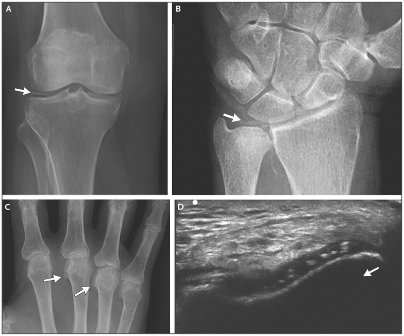
Figure 2. Imaging of Chondrocalcinosis in Patients with CPPD Disease.
Panel A shows a radiograph of a knee with meniscal chondrocalcinosis (arrow). Panel B shows a radiograph of a wrist with chondrocalcinosis of the triangular cartilage (arrow). Panel C shows a radiograph of a hand with hooklike osteophytes (arrows). Panel D shows an ultrasonographic image of a right knee, which was obtained with the transducer in the anatomical axial plane, with the knee flexed 90 degrees. The probe was pointed at the femoral cartilage on the “V” of the patellar groove. Chondrocalcinosis is seen in the substance of the cartilage; the arrow indicates the direction of the probe.

